Decoding Cellular Immunity: Granulocyte and Monocyte Phagocytic Function in Whole Blood
Understanding Phagocytosis in Whole Blood
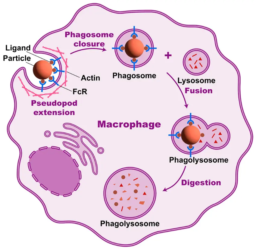
Phagocytosis involves the recognition, internalization, and degradation of particles such as bacteria, fungi, and cellular debris. This complex process is driven by specialized phagocytes—primarily neutrophils (a major type of granulocyte) and monocytes. Both cell types circulate in the bloodstream and respond rapidly to infection or injury.
Unlike isolated cell assays, whole blood phagocytosis assays preserve the native cellular environment, allowing interactions between immune cells, serum components, and plasma proteins. This provides a more accurate representation of in vivo immune activity, making whole blood testing valuable in clinical immunodiagnostics, infection monitoring, and immunotoxicology studies.
Granulocytes: Rapid Responders
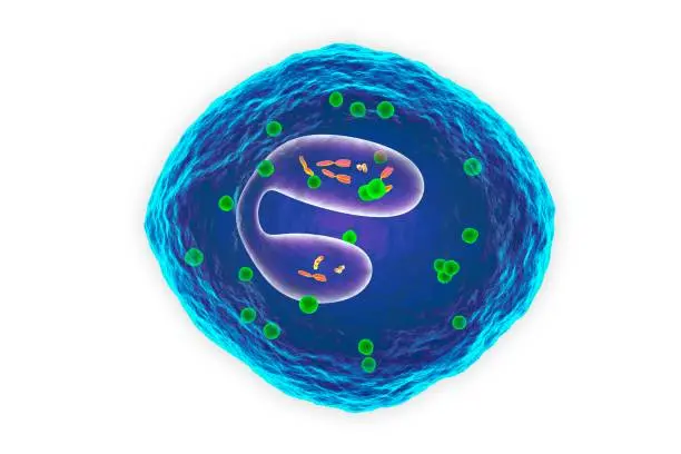
Granulocytes, especially neutrophils, are the most abundant white blood cells in circulation. Their cytoplasm contains granules rich in antimicrobial proteins, enzymes, and reactive oxygen species (ROS). Upon activation, neutrophils rapidly migrate to sites of infection, where they phagocytose pathogens and release oxidative bursts and proteolytic enzymes that contribute to pathogen destruction.
In whole blood assays, neutrophils are typically the most active phagocytes and exhibit rapid uptake of fluorescently labeled bacteria or beads, making them ideal markers for assessing immediate immune responses
Monocytes: Versatile Phagocytes
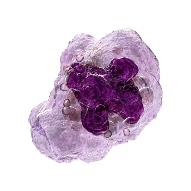
Monocytes are circulating precursors of macrophages and dendritic cells. They are capable of sustained phagocytic activity and contribute to both antigen presentation and cytokine release. While slower to respond than granulocytes, monocytes play a critical role in regulating inflammation and initiating adaptive immunity.
In flow cytometry-based assays, monocytes can be distinguished by CD14/CD16 expression and are evaluated for their ability to internalize particles over extended incubation periods, often revealing delayed but sustained phagocytic engagement.
Methods for Assessing Phagocytic Function